Spinning Bacteria
Words by
Kamal Nahas
Bacteriophages, or “phages” for short, are, without contest, the most successful pathogens on Earth. 1031 of these bacteria-infecting viruses inhabit the planet at any one time. Every time a phage infects a bacterium, the unwitting host makes hundreds more progeny, which liberate themselves by bursting through the bacterial membrane — a grim end called lysis.
Although phages were first discovered in 1917, and the first images of them were obtained as early as 1939, scientists did not fully understand how these viruses infect and lyse their hosts until the turn of the 21st century.
By this time, researchers had narrowed in on a group of phage proteins called holins and endolysins that seemed to be critical for lysis. Holins blow holes in the bacterium’s membrane, while the endolysins make pores in the tough peptidoglycan cell wall surrounding it. Then, the phages flee through the openings. However, many questions remained. Perhaps the biggest of all was, “How do phages time the moment of lysis to maximize their success?”
As a thought experiment, one might think it's best for phages to rupture their hosts early so that their progeny gain a head start on infecting nearby bacteria. These “early” phages could infect more cells sooner, which could yield larger phage populations in the long run, assuming bacteria are abundant and in easy reach. On the other hand, it may be advantageous for phages to remain in the host longer and make more progeny, especially if bacteria are scarce, as this will increase the odds that at least one descendent phage will infect a new host.
Studies in the 1970s suggested that phages might time their escape by sensing the host cell’s electrical gradient. When researchers treated bacteria with cyanide, a poison that disrupts electrical currents, they observed that phage-induced lysis happened earlier than normal. This pointed at a connection between membrane potential and the activation of holins, the proteins that start lysis. And since bacterial cells rely on their membrane gradient to generate ATP, the main energy currency of the cell, this raised the possibility that phages might be using the host’s energy status as a cue for when to burst out.
What remained unclear, though, was whether the host’s electrical gradient (and thus its ATP production) fades gradually over the course of an infection or collapses all at once just before lysis.
If the electrical gradient gradually disappears during an infection, less and less ATP will be available for energy-hungry steps in the phage lifecycle, such as DNA replication or packaging the genome into capsids. Lysis would be an unpredictable process — some phage infections might “burn out” early just because the host cell’s energy ran out. If, however, the electrical gradient collapses suddenly before lysis at the end of the phages’ lifecycle, neither bacterial metabolism nor phage replication would stall in this way, thus maximizing the number of viral progeny. Measuring the electrical gradient over time in infected cells, then, would finally help scientists understand how phages time their escape.
A group of biophysicists at Texas A&M (Angelika Gründling, Michael Manson and Ry Young) finally answered these questions using a clever experiment that involved spinning bacteria. Their results, published in 2001, resolved the mechanistic debate about how phages control the timing of lysis.
{{signup}}
Here’s what they did: First, to measure the electrical gradient across a single bacterium’s cell membrane, they used an unlikely tool: the flagellum. This tiny, whip-like tail — just 20 nanometers thick — spins like a propeller to drive the bacterium forward. The flagellum is directly powered by the cell’s membrane potential; the larger this membrane potential is, the faster the flagellum spins.
Whereas a single flagellum is too small to see under a light microscope, the E. coli cell it is attached to is not. So the scientists had a brilliant idea: Using antibodies, they affixed the bacterium’s flagellum to a glass slide. As the flagellum spun, it remained stationary while the cell itself rotated. Under a basic light microscope, each bacterium spun around in little circles and thus became a visible proxy for the electrical charge across its membrane.1
Once the cells were tethered and spinning, the Texas team’s next focus was timing. If the researchers simply added phages to the bacteria, each infection would begin at a different moment; some phages would attach to hosts right away, while others would take longer. That randomness would make it impossible to tell exactly when lysis occurred, or what caused it.
So instead of using real phages, the researchers engineered each bacterium with a plasmid encoding the genes for holin and endolysin — the two proteins that break open the cell. They put those genes under the control of a sugar-sensitive promoter. That way, they could trigger all the cells to begin making the lysis proteins at once by adding sugar to the growth medium.
They filmed the spinning bacteria under a microscope. Then, they added sugar and started a stopwatch. Across 15 cells, the average time to lysis was 64 minutes. None of the cells burst more than seven minutes earlier or later than this average time.
The researchers also tracked how fast each cell spun, which gave a live readout of the electrical gradient across the membrane. If holins gradually leaked charge, the cells would slow down over time. But they didn’t. The rotations stayed steady right up until the end, then stopped suddenly. And once a cell stopped spinning, it burst about 19 seconds later, on average.
Video: Rod-shaped E. coli bacteria spin in circles around their anchored tails until they rupture and die. Jump to 1:17 and watch the spinning cell on the left. By 1:21 it suddenly slows to a stop, and at 1:38 it bursts. Credit: Gründling A. et al. (2001).
These findings suggested two things: First, that phages carefully time lysis, potentially allowing an opportunity for the formation of sufficient progeny. And second, that phages don’t disrupt the charge gradient until the last moment, which avoids hampering the cell’s metabolism and thus their own replication.
While this seems like strong evidence that phages preserve the cell’s energy until moments before lysis, it was unclear whether this finding would replicate across other phages and experiments. Young’s team had focused exclusively on holins and endolysins, leaving open the possibility that other phage components might disrupt the cell’s charge earlier in the infection.
So next, they wanted to figure out how much of the membrane’s charge could be lost before holins activate. This matters because it sets a boundary: If other phage components interfere with the cell’s electrical gradient — say, by puncturing the membrane or using up energy — they need to stay within that boundary or risk triggering lysis before sufficient phage copies are made.
To determine this limit, the researchers used dinitrophenol, a chemical that drains the membrane’s charge. They added it to spinning cells in gradually increasing doses, watching how long it took for lysis to occur. At low doses, there was a delay; the cells kept spinning for a while before bursting. The more dinitrophenol that the researchers added, though, the shorter the delay became. When the chemical cut the spin rate in half, lysis happened immediately. In other words, holins get triggered when about 50 percent of the membrane’s charge is depleted.
But why are holins sensitive to membrane charge in the first place? The Texas trio argued that, perhaps, it is an evolutionary safeguard for the phages. E. coli plays host to many phage species (Lambda, Mu, T4, and T7, to name a few), but if multiple phages infected the same cell, they would compete for resources and their genes may counteract each other, preventing them from forming viable progeny.
If a second phage tries to invade a cell that’s already replicating another type of phage, it would temporarily disrupt the membrane potential as it injects its DNA into the cell. That small dip in charge could trigger the holins, causing the cell to lyse early. The first phage would thus sacrifice a few progeny to block replication of the newcomer.
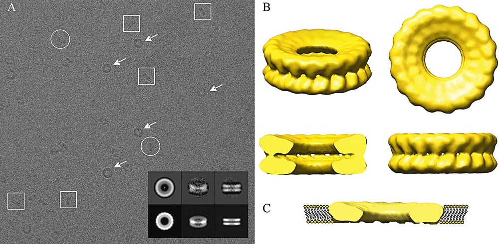
In the decades since this paper was first published, researchers have figured out more structural details about how holins operate. Some have used 2D cryo-electron microscopy, for example, to reveal that these proteins polymerize into a ring-shaped channel with a diameter of nine nanometers in the inner bacterial membrane, allowing endolysin to pass through and break down the cell wall. Using 3D cryo-electron tomography, Young’s team later found that each holin punches two to three large holes in the inner membrane of E. coli, each about 340 nanometers wide — spacious enough for 60-nanometer-thick Lambda phages to escape.
Today, Young’s team could choose other techniques to answer their original questions. There are now charge-sensitive fluorescent proteins, such as pHluorin, whose color changes according to a cell’s electrical gradient. But in 2001, these scientists had little more than a videotape recorder, a stopwatch, a glass surface, and some biophysical ingenuity. Their study reminds us that sometimes simple methods can clarify complex mysteries.
{{divider}}
Kamal Nahas is a researcher-turned-journalist based in Oxford, UK, who covers stories in biology, health, and technology.
Cite: Nahas, Kamal. “Spinning Bacteria.” Asimov Press (2025). https://doi.org/10.62211/83er-59pk
Lead image by Ella Watkins-Dulaney.